

Gingival recession, or dental loosening, is a condition characterized by the loss of marginal tissue. This phenomenon alters the aesthetic appearance of the face and the protective function of the supporting tissues of the tooth.
Located at 8 Boulevard de la Madeleine (Paris 9th), the Madeleine Dental Practice integrates advanced protocols in dentistry and surgery to diagnose the origin of these lesions and implement appropriate treatments. Prompt intervention limits tissue damage and stabilizes the surrounding area of the tooth.
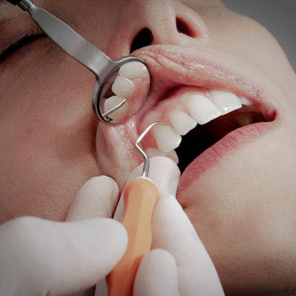
Anatomically, this pathology corresponds to a downward movement of the gingival toward the root. The gingival margin then shifts below the amelo-cemental junction. This tissue displacement results in the direct exposure of the root surface to the oral cavity environment.
The dental crown is protected by enamel, the hardest tissue in the human body. The root, on the other hand, is covered by cement, which is much less dense and therefore more fragile against abrasion, erosion, and acid attacks. The loss of attached gingival , made up of dense keratinized tissue, deprives the tooth of its mechanical barrier against bacterial and traumatic aggression.
It should be noted that this tissue destruction process is irreversible. The gingival has no self-regeneration capacity. Its loss necessitates an assessment, which is the only condition to consider repair through plastic tissue surgery.
At first, gingival recession generally progresses without symptoms. Its slow and silent progression often delays the first consultation. However, when attachment loss intensifies, specific clinical signs appear and become easily identifiable.
The exposed root favors noticeable cervical notches detectable by probing. They are due to either abrasive toothbrushing or excessive occlusal stress (abfraction).
The irregular root surfaces complicate plaque control. This leads to localized gingivitis, with redness, swelling, and bleeding during brushing or probing.
The exposure of the dentin reveals the dentinal tubules. Thermal or chemical variations cause fluid movement that stimulates the pulp nerves and triggers sharp and brief pain.
The gingival retreat visually elongates the crown and creates asymmetry of the necks. This imbalance disrupts the harmony of the smile and becomes particularly bothersome in the anterior zone.
Gingival recession can be caused by various factors. Any intervention should begin with a precise analysis of these elements to effectively guide the treatment strategy.
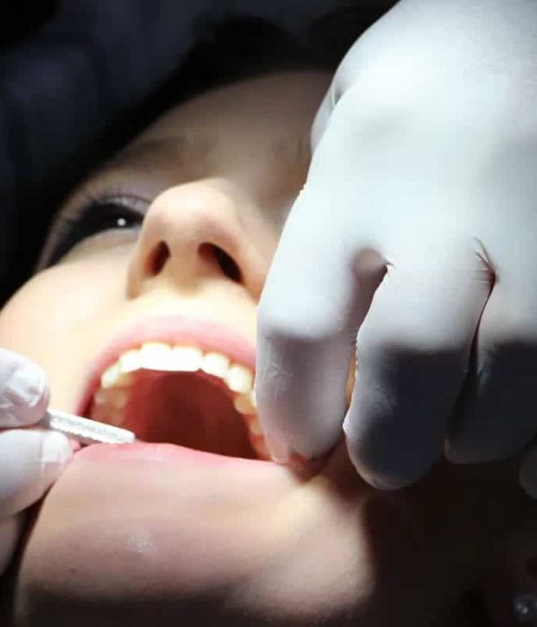
Brushing too hard, especially with horizontal movements, can damage the gingival . The use of hard brushes or abrasive toothpaste with a high abrasiveness index (RDA) exacerbates this phenomenon.
A thin Gingival and low bone thickness increase the risk of recession. Misaligned teeth or certain mucosal frena exert continuous traction that promotes gingival retreat.
Plaque and tartar trigger an inflammatory reaction. This response progressively destroys the supporting tissues and leads to attachment loss as well as resorption of the alveolar bone.
Poorly controlled orthodontic treatment can weaken the bone and cause recession. Tobacco, by reducing vascularization, and oral piercings, through their repeated micro-traumas, are also aggravating factors.
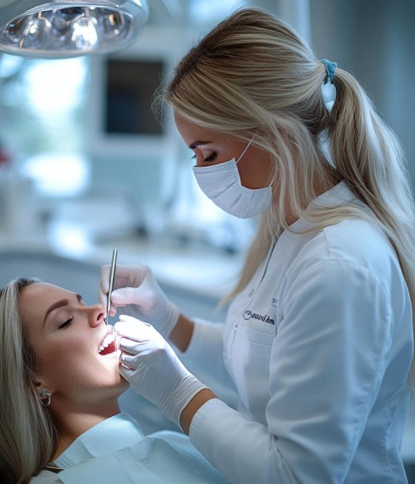
The diagnostic evaluation carried out at the Madeleine Dental Practice is based on standardized medical and radiographic protocols that ensure a high level of precision. During the consultation, the practitioner conducts a thorough examination of the supporting tissues of the tooth.
A calibrated periodontal probe is used to
Gingival recessions are classified according to the Cairo classification (RT1, RT2, RT3). This classification compares the loss of interdental attachment to the vestibular loss in order to evaluate the prognosis for root coverage after treatment.
This clinical examination is supplemented by appropriate radiographic imaging. A panoramic radiograph or three-dimensional imaging of the Cone Beam (CBCT) type allows for assessment of the interproximal bone condition, observation of the thickness of the vestibular bone table, and analysis of the morphology of the bone defect with volumetric precision.
Our intervention primarily aims to control the cause through interceptive therapy, followed by correcting anatomical defects through surgical therapy.
We begin by eliminating the causes of gingival recession. The dentist helps the patient adopt a gentler brushing method to avoid irritating the gingival . It is generally recommended to use the rolling technique and a soft-bristled toothbrush to protect the gingival tissues.
In cases of inflammation related to bacterial biofilm, periodontal cleaning is performed. This treatment includes subgingival scaling and root planing under local anesthesia. It helps to remove bacterial deposits beneath the gingival , clean the surfaces of the tooth roots, and promote healing of the supporting tissues.
The micro-surgery techniques used aim to recreate a dense and stable tissue environment, promoting rapid revascularization.
A connective tissue sample is taken from the donor site, most often the palate. This graft is placed beneath the gingival located near the recession, using a flap displaced towards the crown or a tunneling technique. This method generally allows for good root coverage and a natural aesthetic result with good integration of the grafted tissue.
This technique allows for strengthening the supporting tissues and limiting the progression of gingival recession. The primary objective is to create a more resistant gingival zone to protect the root, even if complete coverage is not always achieved. The healing of the graft occurs progressively through the formation of new blood vessels (angiogenesis).
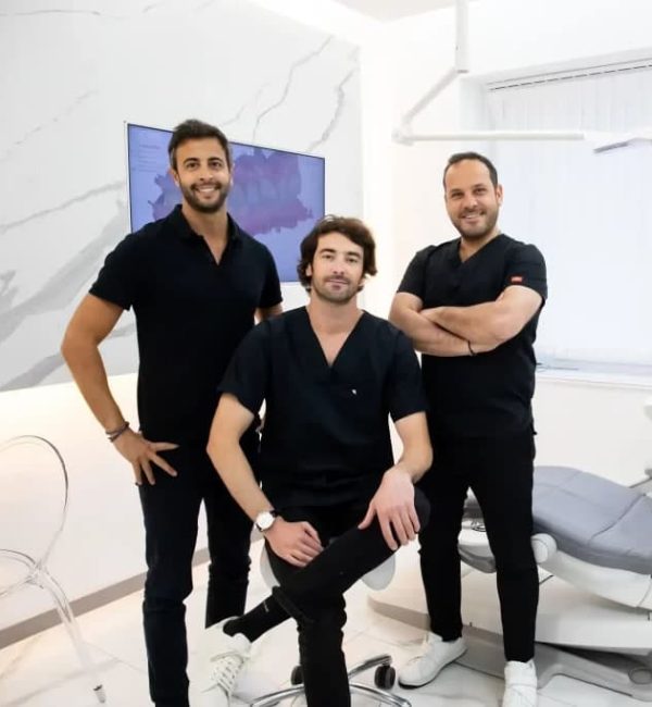
The surgical treatment of mucogingival defects requires great precision and adherence to strict microsurgical protocols. Located in the heart of Paris (Madeleine, 9th arrondissement), the Madeleine Dental Practice uses current scientifically validated techniques in periodontal surgery and tissue engineering.
The medical team, led by Dr. Ralph Badaoui, emphasizes a minimally invasive approach to preserve the tissues as much as possible. Utilizing modern equipment and high-precision microsurgery instruments, the procedures are performed safely and reliably while minimizing postoperative complications.
The health of the teeth directly depends on the quality of the supporting tissues. In case of signs of gingival recession or attachment loss, a clinical evaluation allows for determining the most appropriate treatment plan. The Madeleine Dental Practice, located in Paris 9th, offers a comprehensive diagnosis and personalized treatment based on the characteristics of the gingival tissue.
No. The gingival does not naturally regrow when the root is exposed. Only appropriate management, sometimes surgical, can repair the defect and improve gingival attachment levels.
It is advisable to use a low-abrasive toothpaste (RDA below 50) so as not to further weaken the root. Formulas containing desensitising agents, such as potassium nitrate, help to reduce sensitivity to hot, cold or sweet foods and drinks.
The superficial gingival heals within 7 to 14 days, after which the practitioner will remove the stitches if they are not absorbable. Healing of deeper tissues, such as bone or the attachment of the gingival to the root, continues over a period of 3 to 6 months.
The procedure is performed under local anesthesia and is not painful during the treatment. After the operation, moderate discomfort may occur, especially at the palate if a sample has been taken, but it is generally well managed with standard pain relievers.
Yes, provided you use a soft-bristled head and avoid applying too much pressure. It is not the electric technology that poses a problem, but the force exerted on the gingival, which can promote recession.
Yes, orthodontic treatment remains possible if the condition of the tissues is stable and without inflammation. In some cases, a gingival graft may be performed before orthodontics to strengthen the gingival and limit the risk of exacerbating the recession.